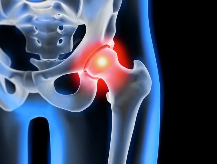
Every year, more and more people are concerned about diseases of the musculoskeletal system, and their development at a young age is becoming more and more observed. This is facilitated not only by lifestyle changes, but also by an increase in the level of interrelated injuries. One of the most common pathologies of the musculoskeletal system is osteoarthritis of the hip joint, characterized by progressive pain and limited mobility. Finally, the disease can lead to complete joint immobility and disability. Treatment of osteoarthritis should be started as early as possible to avoid such undesirable consequences. If it can be stopped conservatively in the early stages of development, then it is possible to restore the function of the hip joint during severe changes and eliminate excruciating pain only with the help of high-tech surgery.
What is osteoarthritis of the hip joint
Osteoarthritis of the hip joint is a chronic degenerative-dystrophic disease in which the hip joint is gradually destroyed. At the same time, all its components are gradually involved in the pathological process, but especially the hyaline cartilage is affected, which leads to narrowing of the joint cavity and deformation of its other components. More often, pathological changes occur in only one hip joint, although both can be affected at the same time.
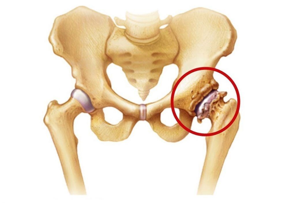
The hip joints are the largest in the human body because they carry the most load during the day. Each is formed by the head of the femur and the acetabulum, which is a pelvic-shaped depression in the pelvis. Both surfaces are covered with smooth, medium elastic hyaline cartilage. This is what ensures the smoothness and unobstructed movement of the head of the femur in the natural depression, and thus allows you to move in different planes.
The movement of the hip joint is provided by a group of muscles connected to it by the fascia. It is also surrounded by ligaments, whose functions are to limit mobility within physiological limits and to ensure the stability of position.
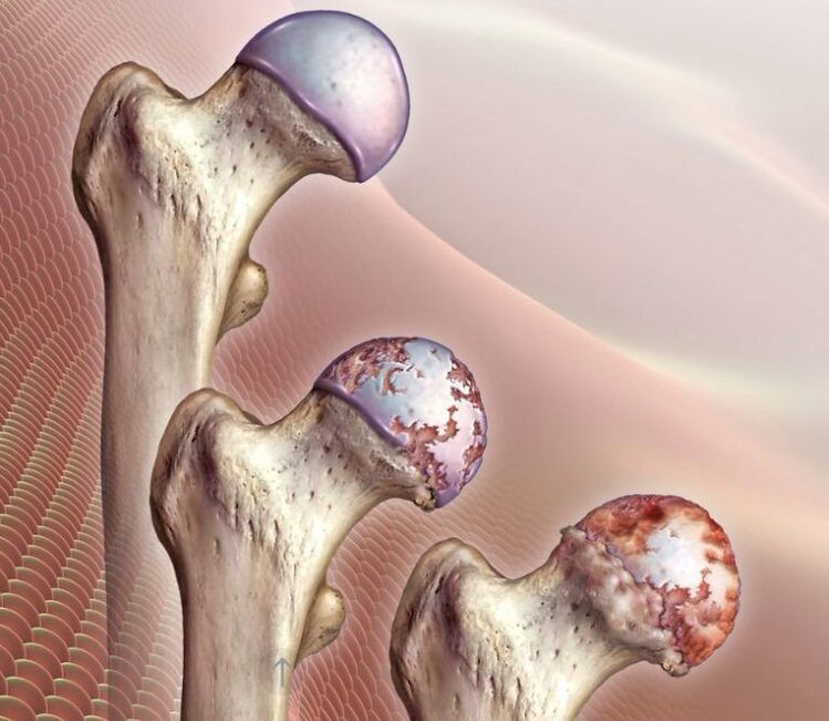
The entire joint is surrounded by an articular capsule covered with a synovial membrane. Its main function is the synthesis of synovial fluid, which lubricates the adjacent parts of the hip joint and at the same time acts as a carrier of nutrients for it. From the synovial fluid, the hyaline cartilage, which covers the head of the femur and the surface of the acetabulum, constantly receives components for the formation of new cells, ie regeneration. This is extremely important for cartilage formation, as it wears out with every movement of the hip, but is normally restored immediately. However, this does not happen when damaged or under the influence of other factors, which leads to the development of osteoarthritis of the hip joint, ie the thinning and destruction of hyaline cartilage.
As a result, ideally deformed areas of smooth cartilage are formed, which increase with the development of pathology. As it wears out, the surfaces of the bones that make up the joint remain exposed. When they come in contact, there is a characteristic crisis and severe pain. This causes osteophytes to form, and in the final stages of development, the head of the femur fuses completely with the acetabulum, making any movement of the femoral joint impossible.
At the same time, osteoarthritis of the hip joint can lead to the development of various inflammatory processes within the joint, including:
- bursitis - inflammation of the synovial sac;
- tendovaginitis - an inflammatory process in the lining of the tendons of the muscles;
- tunnel syndrome - compression of nerves, causing pain radiating along the suffocated nerve.
Reasons
One of the common causes of the development of osteoarthritis of the hip joint is mechanical damage, not only direct injuries, but also micro-damages caused by the destructive effects of excessive loads. One of the most common causes of the disease is a fracture of the neck of the femur.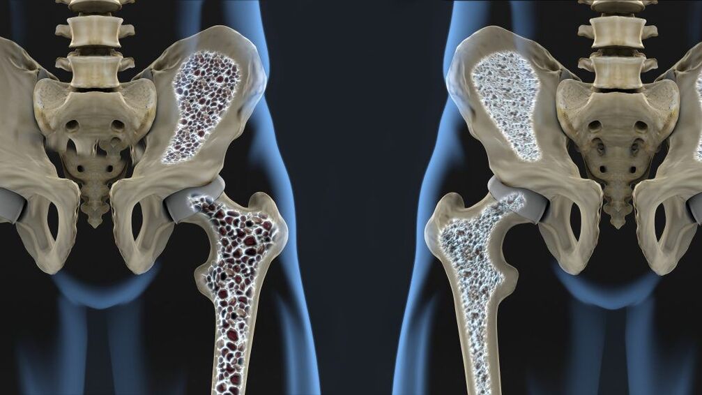 It separates from the femur at an angle of 120 ° and connects it to the head. The presence of osteoporosis significantly increases the likelihood of a hip fracture, but such an injury can also lead to a car accident, falling from a height, a blow, etc.
A fracture of the femoral neck may be accompanied by aseptic necrosis of the femoral head, which will contribute to the development of degenerative-dystrophic changes in the joint. Dysplasia or subluxation of the femoral joint, rupture of its ligaments, transcondylar fractures or fractures of the acetabulum also create favorable conditions for damage to its structures. In such cases, post-traumatic osteoarthritis of the hip joint is diagnosed.
Often, post-traumatic hip osteoarthritis occurs in professional light and heavy athletes, paratroopers, weightlifters, and skaters.
The development of osteoarthritis of the hip joint after injury is associated with impaired compliance (comparability) of articular surfaces, reduced quality of blood supply to the joint components and long-term immobilization. Prolonged inactivity not only worsens blood circulation in the stable area, but also shortens the muscles and reduces their tone. When left untreated or untreated, the likelihood of post-traumatic osteoarthritis increases significantly, leading to the preservation of defects of varying severity. Also, the risks of its development are very intensive, late or, conversely, early, including joint premature overload and inadequate exercise therapy.
Sometimes the disease occurs after surgery on the hip joint due to the formation of scars and additional tissue trauma. However, in some cases, surgery is the only way to eliminate the consequences of the injury.
Excessive loads can also cause changes in the hip joint, as they cause microtrauma. Regular tissue damage activates the division of chondrocytes (cartilage tissue cells). This is accompanied by an increase in the intensity of production of cytokines, which are normally produced in small quantities. Cytokines are mediators of inflammation, in particular the cytokine IL-1, which causes the synthesis of special enzymes that destroy the hyaline cartilage of the hip joint.
In addition, high loads can cause micro-fractures of the subchondral plate. This causes it to gradually shrink and bone growths called osteophytes form on the surface. They can have sharp edges and cause more damage to the joint, as well as damage to the surrounding tissues.
The subchondral plate is the extreme part of the bone that is in direct contact with the hyaline cartilage.
In some cases, it is not possible to determine exactly what causes the development of degenerative-dystrophic changes in the hyaline cartilage of the femoral head and acetabulum. In such cases, idiopathic or primary osteoarthritis of the hip joint is diagnosed.
It has been established today that its development trend may be hereditary, ie. The presence of this pathology in close relatives significantly increases the chances of developing osteoarthritis of the hip joint. It is likely that it has a polygenic inheritance, meaning that its development depends on the presence of many genes. Each of them creates mild conditions for the development of the disease individually, but when combined, it becomes a matter of time, especially when leading a sedentary lifestyle and obesity or, conversely, heavy physical labor.
There is a theory that osteoarthritis of the hip joints is the result of a congenital or acquired mutation of the type II procollagen gene.
There is also secondary arthrosis of the hip joint, which develops against the background of concomitant diseases and age-related changes.
Symptoms
The disease is characterized by pain in the hip joint, limited mobility and the occurrence of a crisis, the severity of which directly depends on the degree of indifference to pathological changes. In the later stages of development, shortening of the affected leg and complete immobility of the hip joint can be observed, which is due to the complete integration of the bone structures that make it up.
At first, the disease can go on without any obvious symptoms and can cause mild, short-term pain. As a rule, they appear after physical exertion, especially after walking, carrying heavy loads, squatting, bending. But as the degenerative-dystrophic changes in the joint progress, the pain intensifies. Over time, they not only become more intense, but also last longer, and the interval between the onset of physical activity and their appearance decreases. At the same time, rest, even for a long time, may not bring relief. Later, pain, for example, after a night's sleep, can afflict such a person with prolonged inactivity of the hip joint.
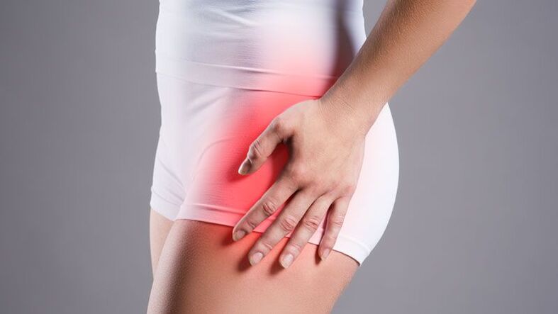
If intraarticular structures damage nearby nerves, pain can spread to the groin, hips, thighs, and knees. However, they tend to be exacerbated by hypothermia. In the final stages of the disease, the pain becomes unbearable. This leads to an unconscious desire to get up and put less stress, which leads to lameness.
Another sign of hip arthrosis is a decrease in range of motion. Most often there is a limitation in the ability to turn the foot in and out, to lift the leg bent at the knee to the chest. Over time, a condition called morning stiffness develops, which disappears after the patient's "sabotage". Later, a compensatory curvature is possible in the pelvis, which causes a change in gait. In the future, patients will completely lose the ability to perform certain movements with the affected foot.
If osteoarthritis of both thighs develops at the same time, the development of the so-called duck walk, in which the pelvis is retracted and the body is bent forward, is observed.
All this can be accompanied by the formation of edema in the hip joint. But when they are overweight, they can be overlooked.
Often, during movements, especially extensors, a crisis occurs in the affected joint. This is the result of exposure to the bony surfaces of the femoral head and acetabulum and their friction with each other. In this case, there is a sharp increase in pain.
In addition, with osteoarthritis of the hip joint, painful spasms of the femoral muscles can occur. With highly advanced degenerative-dystrophic diseases, when the joint space is almost completely gone and the femoral head begins to straighten, a shortening of the affected joint by 1 cm or more is observed.

In general, there are 3 degrees of osteoarthritis of the thigh joint:
- Grade 1 - the joint space of the hip joint narrows and the edges of the bone structures are slightly pointed, which indicates the beginning of the formation of osteophytes. Clinically, there is a slightly pronounced pain syndrome and some movement limitations.
- Grade 2 - the joint space narrows by more than 50%, but less than 60%. Significant osteophytes, as well as signs of cysts in the epiphyses of the bones are observed. Patients report significant limitations in movement in the hip joint, the presence of a crisis during movements, pain and atrophy of the thigh muscles of varying severity can be observed.
- Grade 3 - the joint cavity is reduced by more than 60% or completely absent, and osteophytes occupy a large surface area and are large in size, subchondral cysts are observed. The hip joint hardens, the pain can become unbearable.
Diagnostics
The appearance of pain and other symptoms characteristic of osteoarthritis of the hip joints is a reason to contact an orthopedist. Based on the information obtained during the interview and examination, the doctor may suspect its presence, especially if he has suffered a hip or pelvic injury in the past.
Osteoarthritis of the hip joint is characterized by pain, the intensity of which increases over several years. Less frequently, there is a rapid development of degenerative-dystrophic changes, from a few months to the onset of the first symptoms to severe persistent pain syndrome. It is characterized by increased pain when standing or performing physical activity. Also, for osteoarthritis, morning stiffness lasting up to half an hour is typical and occurs even after prolonged inactivity. Gradually, in the later stages of development, there is an increase in mobility limitations and deformity of the hip joint, which may be noticed by the orthopedist on examination.
However, all patients are necessarily prescribed instrumental research methods, which will be able to confirm the presence of osteoarthritis and determine its degree, as well as to distinguish it from some other diseases accompanied by similar symptoms. As a rule, diagnostics is carried out using the following:
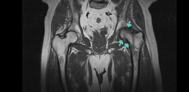
- Radiography - allows to detect the main symptoms of osteoarthritis, in particular, the narrowing of the joint space and the presence of osteophytes. However, in recent years, CT has become a more informative research method, which allows you to more accurately assess the condition of the hip joint.
- MRI is a highly informative method for diagnosing various changes in the condition of soft tissue structures, including cartilage, which allows the detection of the slightest signs of hyaline cartilage degeneration.
Also, patients are given laboratory tests, including KLA, OAM, biochemical blood test, etc. can be assigned. They are required to identify the accompanying diseases that created the initial conditions for the development of secondary osteoarthritis of the thigh joint.
Non-surgical treatment of osteoarthritis of the hip joint
Treatment of degenerative-dystrophic changes in the hip joint with conservative therapy is possible only with first and second degree osteoarthritis. The prescribed measures can improve the patient's condition, stop or at least slow down the course of the pathology, and thus maintain the ability to work. However, together they do not lead to a complete regression of the changes that have already taken place.
Today, as part of conservative treatment of osteoarthritis of the hip joint, the following are prescribed:
- drug treatment;
- exercise therapy;
- physiotherapy.
Patients are also advised to make some lifestyle adjustments. Thus, when you are overweight, it is worth taking measures to reduce it, that is, to increase the level of physical activity and reconsider the nature of nutrition. It is recommended to reduce the intensity of exercise if the patient is actively engaged in sports and the joint is loaded, which causes microtrauma.
Medical therapy
Drug treatment for osteoarthritis of the hip joint is always complex and includes drugs of different groups aimed at reducing the severity of symptoms of the disease and improving the flow of joint metabolic and other processes. O:
- NSAIDs - drugs with anti-inflammatory and analgesic effects, produced both orally and in the form of topical agents, allow you to choose the most effective and convenient option for use;
- corticosteroids - drugs that have strong anti-inflammatory properties and are often used in the form of injectable solutions, as they cause the development of undesirable side effects when choosing systemic therapy;
- chondroprotectors - drugs synthesized on the basis of natural components of cartilage tissue used for the regeneration of the body (prescribed for long courses);
- muscle relaxants - drugs prescribed for muscle spasms that cause pain of varying severity;
- B vitamins - help improve nerve conduction required for the development of carpal tunnel syndrome;
- Drugs that improve microcirculation - help to increase the intensity of blood circulation in the affected area, which leads to an increase in the rate of metabolic processes and helps to restore damaged cartilage.
If concomitant diseases are detected, appropriate specialist consultation and appropriate treatment are indicated.
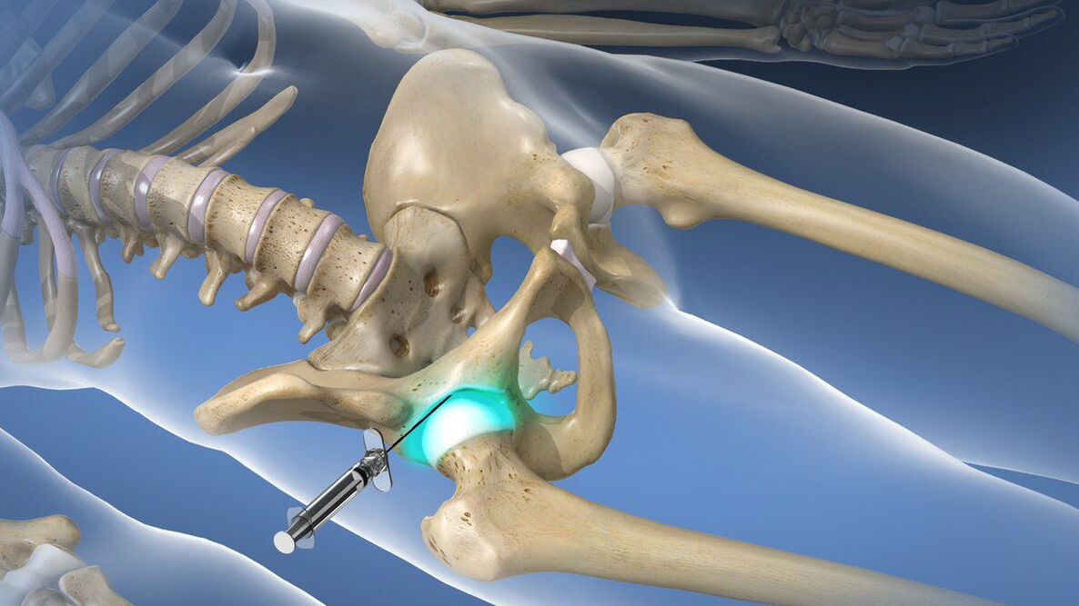
With very severe, debilitating pain syndrome that cannot be relieved with prescribed NSAIDs, intraarticular or periarticular blockades may be performed. They include injection of local anesthesia in combination with corticosteroids directly into the joint cavity, which leads to a rapid improvement in well-being. However, such procedures can be performed only by a qualified specialist in a medical institution, otherwise the risk of complications is high.
exercise therapy

Physiotherapy exercises play a leading role in the non-surgical treatment of osteoarthritis of the hip joints in both idiopathic and post-traumatic forms. However, a number of exercises should be selected individually, taking into account the nature of the previous injury, the patient's level of physical development and existing concomitant diseases.
Exercise therapy should be done every day in a comfortable environment without haste. All movements must be performed smoothly without shaking to avoid damaging the already deformed hip joint. This will allow:
- reduce the intensity of pain syndrome;
- increase joint mobility;
- reduce the risk of muscle atrophy;
- increase the intensity of blood circulation and metabolic processes.
Physiotherapy
To increase the effectiveness of the prescribed measures, it is often recommended that patients with osteoarthritis of the hip joint undergo a course of physiotherapy procedures. Traditionally, those with anti-inflammatory, anti-edematous and analgesic effects are selected. O:
- ultrasound therapy;
- electrophoresis;
- magnetotherapy;
- laser therapy;
- shock wave therapy, etc.
In some cases, plasmolifting, ie the application of the patient's own blood plasma, which is purified and saturated with platelets, is indicated. To obtain it, venous blood is taken and then centrifuged. As a result, it is divided into erythrocyte mass and plasma, which are used to treat degenerative-dystrophic changes in the hip joint.
Surgery for osteoarthritis of the hip joint
When grade 3 hip arthrosis is diagnosed, patients are referred for surgery. It can also be performed with the ineffectiveness of conservative therapy and persistent pain and limited mobility in stage 2 of the disease.
In general, the instructions for hip surgery are as follows:
- a significant reduction in the size of the joint cavity;
- persistent, severe pain;
- significant movement restrictions.
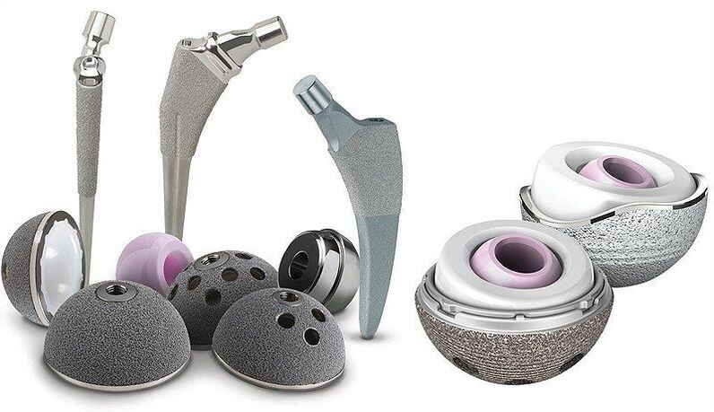
The most effective and safest operation for osteoarthritis of the hip joint is arthroplasty. Today, regardless of the reasons for its development, it is recognized as the gold standard for the treatment of this pathology. The essence of this type of surgery is to replace part of the components of the hip joint or completely artificial endoprostheses. The prostheses themselves are made of biocompatible materials and are durable.
Their installation allows to completely restore the normal mobility of the pathologically altered hip joint, relieve pain and allow the patient to live a full life. The type of arthroplasty for each patient is selected individually based on the degree of destruction of various components of the joint.
The most effective is total or total hip arthroplasty. This involves the replacement of the entire joint with an artificial endoprosthesis, ie the acetabulum, the head and neck of the femur. Such prostheses can serve for 15-30 years without interruption and provide full restoration of joint function.
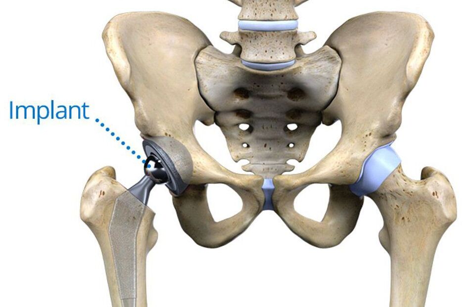
They are installed without cement or with the help of special cement. The first method is more suitable for young patients, because the prosthesis in the pelvis involves attaching its own bone to the sponge layer. For the elderly, even in the presence of osteoporosis, the method of implantation of an endoprosthesis using cement, which holds the artificial material firmly to the bone surfaces, is more appropriate.
If normal hyaline cartilage is preserved on the surface of the acetabulum, patients may be offered partial arthroplasty. Its essence is to replace only the head and neck of the femur with an endoprosthesis. Today there are 2 types of such structures: monopolar and bipolar.
The former are less reliable, requiring total arthroplasty after installation. This is due to the fact that the modified artificial femur is rubbed directly into the cartilage of the acetabulum while the head is moving, which causes it to wear faster.
Bipolar endoprostheses do not have such a disadvantage, because in them the head of the artificial femur is already closed with a special capsule adjacent to the acetabulum. Therefore, the cartilage surrounding it is not deformed, because the capsule serves as a kind of buffer and artificial replacement for the natural hyaline cartilage of the femoral head.
However, postoperative rehabilitation is indicated for all patients, regardless of the type of endoprosthesis performed. It consists of the appointment of drug therapy, exercise therapy and therapeutic massage. Recovery time depends on individual characteristics. However, it should be borne in mind that the effectiveness of the operation directly depends on the quality of compliance with the doctor's recommendations during rehabilitation.
Thus, osteoarthritis of the hip joint is a common disease of the musculoskeletal system that can occur even in the absence of direct preconditions for its development. This pathology can cause not only severe pain, but also disability, so it is important to diagnose and take measures to stop its development, even at the first signs. However, the current level of development of medicine allows to cope with the advanced cases of osteoarthritis of the hip joint and restore the full range of motion in it, as well as to get rid of severe pain forever.


